Medical Imaging "Super Processing": Seeing the Unseen
In the critical field of diagnostic medicine, a diagnosis is only as accurate as the image that informs it. Modern medical imaging devices—MRI, CT, PET, and Ultrasound—are marvels of sensor engineering. However, the onboard computers that process this raw sensor data are often constrained by thermal limits, power budgets, and hardware cycles locked in years before the machine was installed.
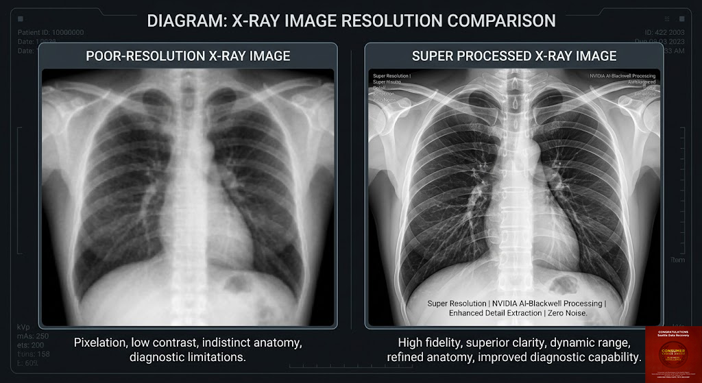
Seattle Data Recovery bridges this gap. Located in the University of Washington Medical Building, we utilize proprietary Super Processing to re-render and enhance medical imaging data. We take what
Medical Imaging "Super Processing": Seeing the Unseen
subtle anomalies that standard processing misses.
The Hardware Gap: Why Your Scanner Is Limited
Even a multimillion-dollar MRI machine does not possess the computational throughput of a dedicated supercomputing cluster. Most medical devices rely on embedded processors or mid-range workstations optimized for stability rather than peak performance. They often lack:
-
The NVIDIA RTX 50 Series ("Blackwell"): The latest generation of GPU architecture designed for massive parallel processing and AI-driven image reconstruction.
-
Terabytes of ECC RAM: The memory capacity required to load entire 3D volumetric datasets into a high-speed buffer without compression or downsampling.
-
Dual Intel XEON CPUs: The raw multi-threaded power needed to solve complex reconstruction algorithms in real-time.
At our laboratory, we process your data on 128-bit hardware stacks that far exceed the specifications of standard OEM consoles. This allows us to apply computationally expensive algorithms—such as iterative reconstruction and deep learning-based denoising—that onboard hospital systems cannot handle.
The "Super Processing" Difference for Critical Modalities
By applying our industrial-grade processing power to medical data, we enhance the diagnostic value of every scan.
1. MRI: Unveiling Soft Tissue Nuance
Magnetic Resonance Imaging relies on "k-space" data to reconstruct an image. Standard machines often use "Fast Fourier Transform" shortcuts to speed up this process, which can introduce noise or blur fine details.
-
Our Enhancement: We use 128-bit precision to re-process the raw data, eliminating the "rounding errors" that obscure micro-structures.
-
Clinical Value: This results in sharper differentiation between healthy tissue and potential tumors in the brain or breast, enabling earlier detection of life-threatening conditions.
2. CT: Reducing Artifacts in 3D Space
Computed Tomography creates massive 3D datasets that require immense processing to visualize without artifacts (like "beam hardening" from bone or metal implants).
-
Our Enhancement: Using the Tensor Cores of the RTX 50 series, we run advanced artifact-reduction algorithms that clarify the interface between bone and soft tissue.
-
Clinical Value: This provides a clearer view of complex fractures, vascular blockages, or internal bleeding, allowing surgeons to plan interventions with sub-millimeter accuracy.
3. Ultrasound: Clarifying the Signal
Ultrasound is prone to "speckle noise" that can make images look grainy.
-
Our Enhancement: We use AI-driven signal separation to distinguish true acoustic reflections from digital noise.
-
Clinical Value: This delivers a "glass-like" clarity for fetal imaging and cardiac assessments, improving real-time detection of valve defects and clots.
Seeing Better Saves Lives
The correlation between image quality and patient outcomes is well-documented. A clearer image means:
-
Faster Decisions: Radiologists spend less time squinting at ambiguous shadows and more time diagnosing.
-
Earlier Intervention: Detecting a lesion when it is millimeters smaller can be the difference between a simple procedure and a complex surgery.
-
Reduced Radiation: By utilizing advanced denoising, we can often extract high-quality images from lower-dose scans, improving patient safety.
Trust the Local Experts
We are not just data recovery specialists; we are partners in patient care. Based in the heart of Seattle's medical community, we provide the secure, HIPAA-compliant processing power that modern medicine demands.
See what your scanner is missing. Contact Seattle Data Recovery to discuss our Medical Imaging Super Processing services.
Here are the sources for the information used in the articles, formatted in MLA style:
References
Photography & Imaging Formats
-
"Adobe Camera Raw." Adobe Help Center, Adobe, https://helpx.adobe.com/camera-raw/using/introduction-camera-raw.html.
-
"Digital Negative (DNG)." Adobe Creative Cloud, Adobe, https://www.adobe.com/creativecloud/file-types/image/raw/dng-file.html.
-
"Fujifilm X RAW Studio." Fujifilm X Series, Fujifilm, https://www.fujifilm-x.com/en-us/products/software/x-raw-studio/.
-
"Imaging Edge Desktop Features." Sony Support, Sony, https://support.d-imaging.sony.co.jp/app/imagingedge/en/instruction/1_1_features.php.
-
"JPEG vs. Raw." Adobe Creative Cloud, Adobe, https://www.adobe.com/creativecloud/file-types/image/comparison/jpeg-vs-raw.html.
-
"Raw Image Format." Wikipedia, Wikimedia Foundation, https://en.wikipedia.org/wiki/Raw_image_format.
-
"TIFF vs. JPEG: A Photographer’s Guide." Richard Photo Lab, https://richardphotolab.com/blogs/post/jpeg-vs-tiff-a-photographers-guide.
-
"What Is a TIFF File?" Shorthand, https://shorthand.com/the-craft/what-is-a-tiff-file/index.html.
Professional Photography & Labor
-
Bossert, Olivia. "Why Your Fashion Photography Portfolio Is the Backbone of Your Career." Olivia Bossert Education, https://www.oliviabosserteducation.com/photography-education/why-your-fashion-photography-portfolio-is-the-backbone-of-your-career.
-
"How Much to Charge for Photo Editing?" Clipping Path Experts, https://www.clippingpathexperts.com/blog/how-much-to-charge-for-photo-editing/.
-
Jiao, Belinda. "How Long Does It Take a Professional Photographer to Edit Photos?" Belinda Jiao Photography, https://www.belindajiao.com/blog/how-long-professional-photographer-edit-photos.
-
Motta, Daniel. "What Happens After the Photo Shoot: An Inside Look at Post-Production." Daniel Motta Photography, https://danielmottaphotography.com/blog/what-happens-after-the-photo-shoot-an-inside-look-at-post-production/.
-
"Why Every Photographer Needs a Portfolio." Fstoppers, https://fstoppers.com/education/why-every-photographer-needs-portfolio-676508.
Medical Imaging & Healthcare Data
-
"Benefits and Risks of MRI." U.S. Food and Drug Administration, https://www.fda.gov/radiation-emitting-products/mri-magnetic-resonance-imaging/benefits-and-risks.
-
"CT Scans Explained: How They Help Doctors Detect and Treat Serious Conditions." Midtown Medical Imaging, https://www.midtownmedicalimagingfortworth.com/ct-scans-explained-how-they-help-doctors-detect-and-treat-serious-conditions/.
-
"Effective Strategies for Data Backup & Recovery in Healthcare." Spin.AI, https://spin.ai/blog/effective-strategies-for-data-backup-recovery-in-healthcare/.
-
"Healthcare Data Storage and Retrieval Challenges." Nexstor, https://nexstor.com/healthcare-data-storage-and-retrieval-challenges/.
-
"How MRI Scans Have Saved Lives." Hollywood Diagnostics Center, https://hdc1984.com/how-mri-scans-have-saved-lives/.
-
"Magnetic Resonance Imaging (MRI)." National Institute of Biomedical Imaging and Bioengineering, https://www.nibib.nih.gov/science-education/science-topics/magnetic-resonance-imaging-mri.
-
"Positron Emission Tomography (PET)." Johns Hopkins Medicine, https://www.hopkinsmedicine.org/health/treatment-tests-and-therapies/positron-emission-tomography-pet.
-
"Ultrasound’s Impact on Healthcare Risk Management." Acertara Labs, https://acertaralabs.com/ultrasounds-impact-on-healthcare-risk-management/.
-
"What Is Medical Imaging?" Intel, https://www.intel.com/content/www/us/en/learn/what-is-medical-imaging.html.